PRP & PROLOTHERAPY FOR SACRO-ILIAC JOINT AND PUBIC LIGAMENT INCOMPETANCE
The physiotherapists at Sydney Spine & Pelvis Centre have been involved in diagnosis of mechanical sacro-iliac joint and pubic ligament incompetence for 17 years. We continue to provide expert advice on appropriate treatment and rehabilitation for such injuries, as well as being able to advise on referral to the best Medical specialists for PRP or prolotherapy at the pubic or sacro-iliac ligaments, or costo-transverse ligaments in the thorax if it is required.
Please note as physiotherapists we do not perform either PRP or prolotherapy injections.
SO WHAT IS PROLOTHERAPY VS PRP?
Prolotherapy treatment for the pubic symphysis and sacroiliac joints has been researched in Sydney, Australia over the past 14 years under the guidance of the Low Back Interest Group (LBIG Australia).
Data from this research has been published: M Cusi, J Saunders, B Hungerford, T Wisbey-Roth, P Lucas, S Wilson (2008) The use of prolotherapy in the sacro-iliac joint. Br. J. Sports Med
PRP injections into the SIJ Interosseous ligament structure
In the past 3 years the use of PRP (Plasma Rich Platelets) derived from the patient’s own blood has been shown to provide greater improvement in function with less pain than prolotherapy (Saunders J et al 2016. A comparison of PRP & hypertonic glucose injections in the treatment of SIJ mechanical incompetence. 9th Interdisciplinary WCLBP P173-4). The biological effects are the same as in prolotherapy but proliferation of collagen is quicker, and often less injections are required to create articular stability. The protocol for assessment, and post injection rehabilitation follows the same guidelines as prolotherapy.
Prolotherapy
Prolotherapy involves the injection of a proliferant solution into a ligament, with the aim to cause proliferation of growth of collagen tissue which is the principle component of ligaments. There are many proliferant solutions, however, in our research we chose glucose as the safest option. The strength of the glucose solution has been researched in order to produce optimal results, that is, to produce collagen as close to normal ligament structure, rather than scar tissue which has been shown to stretch over time.
Biological Effects
The proliferant solution works by creating a cascade of biological events that cause the release of pre-collagen growth factor (PGF) which encourages specific cells, known as fibroblasts, into the injection area. It is the fibroblasts that secrete the collagen tissue. This is a biological process that takes about 6 to 8 weeks to be completed. This explains why people undertaking prolotherapy injections do not immediately feel better, and also why we wait 6 to 8 weeks between injections.
Why would Prolotherapy be indicated?
Prolotherapy may assist collagen growth in ligaments that have been damaged by trauma or injury. At present the joints being treated in this trial are the sacroiliac joint, the pubic symphysis, and the costo-transverse joints.
Prolotherapy in the pelvis
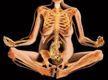
The joints of the pelvis, the sacroiliac joints and the pubic symphysis, are inherently stable joints. That means that, normally, the ligament structure and muscles surrounding the joints support the pelvis so that very little movement occurs, even under large loads such as walking or running. During late pregnancy and labour, the amount of movement occurring at the sacroiliac joints and pubic symphysis will increase due to the effects of hormones such as relaxin. Under normal circumstances, however, movement will return to normal and the ligaments will become firm again within a short time of giving birth.
Pain originating from the pelvic joints, ligaments, and muscles, can be debilitating, and can make it difficult to sit, walk, stand, and even sleep. In most instances, pelvic pain and sacroiliac pain can be successfully treated with manual therapy and specific exercise rehabilitation. Occasionally, the force of the injury (for example due to a nasty fall or motor vehicle accident) may be sufficient to strain the pubic or sacroiliac ligaments. The majority of ligament strains also heal with time, and with appropriate treatment, however in a small percentage of people, the collagen within the ligament remains damaged, and creates ligament laxity. This affects the ability of the pelvis to maintain its stable joint alignment so that weight can be transmitted from the upper body down onto the legs. It is this small group of patients who have increased laxity of the pelvic ligaments that have been shown to respond to prolotherapy (Dorman et al, 1995), when the injection is given under CT Scan guidance (ensures that the proliferant is placed correctly, and where it will have the best effect).
At present, any patients seeking prolotherapy must undergo a complete examination from a therapist fully trained in assessing stability and ligament function. They will determine whether there is any indication that there is significant ligament damage and laxity that may indicate prolotherapy could be of some benefit toward improving function. All candidates for prolotherapy into either the sacroiliac or pubic ligaments must undergo a 3 month trial of specific lumbo-pelvic stability exercises before being accepted into the program, and Sydney Spine & Pelvis has been involved in designing and providing these exercise programs for patients for 15 years. The prescribed exercises improve muscle activation around the lumbar spine and pelvis, and in many cases these exercises alone will improve lumbo-pelvic stability so that the prolotherapy is not needed.
PROLOTHERAPY IS NOT A FORM OF PAIN RELIEF. It may improve pain over time because the ligament structure gets stronger and this improves the ability of the underlying joint to cope with forces placed onto it, for example, when walking. In fact, the injection may initially increase pain. The pain usually settles within 3 to 4 weeks.
In order to maintain the effects of the injections, patients need to continue performing the lumbo-pelvic exercises for a year after intervention, and this should also accelerate functional improvement.
PRP & Prolotherapy and Medication
DO NOT TAKE ANTI-INFLAMMATORY MEDICATION
(Aspirin, Nurofen, Nurofen plus, Voltaren, advil, prednisone, hydrocortisone, other prescription antinflammatories etc). Although they can effectively reduce the pain, they will fight the very effects of the injection! These medications need to be stopped 14 days prior to your first injection.
Pain relieving drugs such as digesic, panadol, panadeine, and tramyl are accepted.
Activities following treatment
You should carry on with your normal activities as much as possible. Do not engage however in high level activity, putting more than the average physical load on your pelvis (running, cycling and weight lifting are not a good idea. Other activities which you do should be checked with the physiotherapist.)
Prospective patients will be provided with more detailed information during their medical consultation.
MEDICAL /PHYSIOTERHAPY TREATMENT PROTOCOL
In order to ensure that PRP or prolotherapy is only provided to patients who have a true ligament insufficiency, a protocol has been created that determines each patients’ appropriateness for treatment. This protocol is based on the work and research of Dr Andry Vleeming, Diane Lee, Dr Jan Mens, Dr Hans Ostgaard, Dr Thomas Dorman and Dr Barbara Hungerford, plus numerous other clinicians who continue to strive for the answers on best medical treatment for lumbo-pelvic injuries.
The protocol outlined here assesses the sacroiliac joint and pubic symphysis only.
All steps in this protocol must be undertaken to determine what the appropriate treatment should be, and whether a diagnosis of Sacro-iliac or pubic insufficiency can be made.
Step 1
The therapist must clear any pubic symphysis or sacroiliac joint articular dysfunctions, such as fixated joint or compressed joint dysfunctions. This assumes that the therapist has therefore received training from either Barbara Hungerford (Physiotherapist, Sydney, Australia), Diane Lee (Physical Therapist, Vancouver /White Rock, BC, Canada), or Trish Wisbey Roth.
Step 2
Assess pubic symphysis function and stability
The pubic symphysis should be assessed to determine if pelvic insufficiency is occurring due to insufficiency of the pubic symphysis ligaments, SIJ ligaments, or both. In order to make a diagnosis of pubic symphysis ligament laxity the following tests will all be positive:
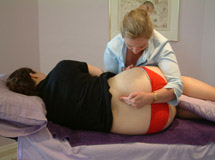
1 The Pubic Shear Test
- PT palpates the superior pubic tubercles with both index fingers
- Ask the patient to lengthen the right leg along the bed, and feel whether pubic alignment remains stable, or whether an inferior translation of the right pubic ramus occurs during this maneuver. Repeat on the left.
- Retest while patient activates their core muscles. Improved?
2 Pubic Passive Joint Glide test
- PT positions the right heel of the hand onto the superior border of the right pubic ramus and heel of left hand onto the inferior border of the left pubic ramus.
- PT creates a translational force of the right pubic ramus inferiorly relative to the left (left supports left ramus), and then of the left ramus superiorly relative to the right.
- Ask patient to gently activate their core muscles (pelvic floor and transversus abdominis). Retest pubic rams translation during activation. All joint translation should be controlled.
- Swap hands so that right heel is on left superior border of the left pubic ramus and heel of left hand onto the inferior border of the right pubic ramus. Retest stability of joint to resist translation.
3 Flamingo View x-rays
Comparing pubic translation while standing on each leg should be performed if pubic instability is diagnosed by the clinical tests. All three tests would need to be positive to consider a provisional diagnosis of pubic instability.
Step 3
Assess sacroiliac joint function and stability
In order to make a diagnosis of SIJ ligament laxity the following tests will all be positive:
- The Stork test on the side of single leg support (Hungerford et al, 2004). The innominate will rotate anteriorly relative to the sacrum as the patient stands onto that leg
- PPPP Test (Posterior pelvic pain provocation test) (Ostgaard et al, 1995). Posterior translation of the innominate in supine lying with the hip in 90 degrees hi flexion may provoke the patients symptoms.
- ASLR Test (Active Straight Leg Raise Test) (Mens et al, 1999, 2000). During an ALSR the patient will describe either difficulty lifting the leg on the affected side, pain, or an inability to maintain the lumbo-pelvic region in neutral during the maneuver.
- ASLR Test with passive compression. Passive compression across either the pubic symphysis or sacroiliac region tends to significantly improve the ability to perform the ASLR (refer to Lee D. 2004).
- Passive SIJ articular glide Test (Lee D. 2004). The therapist will palpate a significant increase in articular translation of the iliac surface relative to the sacral surface in either the A-P or the vertical plane of the joint, on the side of the injury in comparison to the uninjured side. This increase in articular translation will also be present when the therapist moves the joint surfaces in to the closed pack position of the sacroiliac joint (normally there should be no translation palpable in the closed pack position). The increased articular translation on the side of the injury will not be reduced significantly (>50%) with activation of the local lumbo-pelvic stabilization muscles i.e transversus abdominis, lumbar multifidus, and the pelvic floor. (Normally local muscle activation will induce the closed pack position of the sacroiliac joint and minimize any movement at the SIJ). This step assumes that the patient has been taught how to specifically activate the local stabilizing muscles of the lumbo-pelvic region.
- All SIJ tests must be positive to make a provisional diagnosis of an inability to stabilize the SIJ (failed load transfer through the pelvis).
Step 4
Research ash shown that activation of transversus abdominis (TA), lumbar multifidus, and gluteus maximus can increase tension in the posterior layers of Thoraco-lumbar fascia, and thereby increase SIJ stiffness (Barker et al, 2004; Richardson et al, 2002; van Wingerden et al, 2004). The majority of patients who report posterior pelvic pain of musculo-skeletal origin, will improve their ability to stabilize the pelvis during functional tasks by learning how to turn these muscles on and strengthen them. This occurs because improved function of the lumbo-pelvic muscles creates the necessary tension onto the sacroiliac and pubic ligaments, with the consequence of increased articular compression during weight bearing. That means the pelvis is more stable during movement.
The protocol developed by the LBIG group suggests that all clients who have been diagnosed with Failed load transfer through the pelvis undergo a 3 month program of specific retraining for lumbo-pelvic stability. The client must be able to activate the local lumbo-pelvic muscles prior to re-assessment of pelvic stability.
The areas to focus on are:
- isolation of TA, lumbosacral multifidus, & pubococcygeus (the core) in neutral lumbo-pelvic alignment, without substitution of rectus abdominis, the obliques, erector spinae, or coccygeus.
- ability to maintain an isolated activation of ‘the core’ during functional tasks such as sitting, walking, lifting.
- ability to maintain isolated contraction of the core for extended periods (endurance)
- ability to maintain core activation as they progress to exercises that enhance
- gluteus medius activity without substituting TFL or adductors
- gluteus maximus activity without substituting hamstrings
Step 5 Final Diagnosis
After 3 months, reassess the pelvic function and stability as previously described in step 3.
If all tests remain positive, and the ability to activate and maintain core activation does not significantly improve the SIJ passive articular glides, then the test results suggest that force closure from the muscle activation is not being transmitted via the fascia and posterior SIJ ligaments to adequately compress the SIJ and create joint stability. In other words, the positive results suggest that damage to the posterior SIJ ligaments is affecting the force closure mechanism that increases functional stability of the SIJ (Vleeming et al, 1995). In this circumstance, prolotherapy may be indicated to enhance the collagen structure of the affected ligaments, thereby augmenting force closure. CT guided injections of glucose are suggested in order to ensure that the proliferant is specifically released into the affected ligaments.
DISCLAIMER
The accepted Radiological examination used to identify possible damage to the posterior SIJ ligaments and SIJ insufficiency is a SPECT CT scan. This can only be ordered by a medical practitioner. Otherwise, the protocol outlined above is based on conservative palpation techniques and therefore cannot be considered as precise for the determination of which ligament has been damaged.
It is up to the treating therapist to determine if the client has actually performed the exercise program sufficiently to be eligible for reassessment regarding referral to a specialist for either PRP or prolotherapy treatment.


